
The objective is to maintain a safe and secure environment in the research MRI unit for all research participants and personnel.
THE MRI MAGNET IS ALWAYS ON. DO NOT ENTER ZONE 3 AND 4 WITHOUT AUTHORIZATION FROM MRI STAFF.
Zones
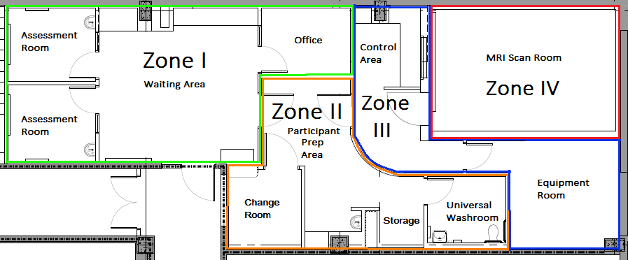
Zone I:
Readily accessible areas by the general public outside MR environment (waiting room)
Zone II:
Area between Zone I and Zone III where individuals are supervised by MR personnel (participant prep area)
Zone III:
Restricted transition area between Zone II and Zone IV that is supervised and controlled by MR personnel (control room and restricted waiting area)
Zone IV:
WARNING: Area controlled and strictly supervised by Level Two MR personnel. This is a potentially hazardous area with presence of very strong magnetic fields. MRI MAGNET IS ALWAYS ON. DO NOT ENTER WITHOUT BEING SCREENED BY/WITH AUTHORIZATION FROM THE MRI TECHNOLOGIST ON-SITE.
Emergency Policies
The MRI Emergency Code Blue and White policy is for all personnel to provide the appropriate response to medical and behavioural emergency situations in the MRI unit.
The MRI Emergency Procedures Fire policy is for all personnel to provide the appropriate response to a Code Red situation in the MRI unit.
The MRI Emergency Procedures Flood policy is for all personnel to provide the appropriate response to a flood emergency situation in the MRI unit
For Scientists
Principal Investigators interested in using MRI as a part of their research study are required to submit a MRI application form with their eREB submission on the eREB platform. Please see the link below for more information:
Scanner: 3.0T Siemens Prisma NUMARIS/4 XA30
v.1.1 | Updated December 2022
Brain Sequences
MR Sequence | Scan Parameters | Scan Time | Purpose | License |
3D T1 MP RAGE (Magnetization Prepared Rapid Acquisition Gradient Echo) | (0.8x0.8x0.8)mm3 , TR=1870ms, TE=3.14ms, Flip Angle=9o, Sagittal FOV =256x250mm2, 240 slices, GRAPPA=2 | 5:03 | High resolution T1 weighted 3D sequences with magnetization preparation for brain morphometry which provides excellent contrast properties for cortical segmentation. | Siemens |
3D T1 MultiEcho MPRAGE (Magnetization Prepared Rapid Acquisition Gradient Echo) | (1.0x1.0x1.0)mm3 , TR=2350ms, TE=1.69/3.55/5.41/7.27ms, TI=1100ms Flip Angle=7 o, Sagittal FOV =256x256mm2, 176 slices, GRAPPA=2 | 5:38 | High resolution T1 weighted 3D sequences with magnetization preparation for brain morphometry which provides excellent contrast properties for cortical segmentation. Higher bandwidth reduces distortions while combining echoes recovers SNR. | C2P Agreement |
3D T1 MPRAGE (Magnetization Prepared Rapid Acquisition Gradient Echo) With Navigator | (1.0x1.0x1.0)mm3 , TR=2500ms, TE=2.88ms, TI=1070ms Flip Angle=8o, Sagittal FOV =256x256mm 2, 176 slices, GRAPPA=2 | 7:12 | High resolution T1 weighted 3D sequences with magnetization preparation for brain morphometry which provides excellent contrast properties for cortical segmentation. Prospective compensation for subject motion using volumetric navigators (vNavs) | C2P Agreement |
3D T1 MultiEcho MPRAGE (Magnetization Prepared Rapid Acquisition Gradient Echo) With Navigator | (0.8x0.8x0.8)mm3 , TR=2500ms, TE=1.86/3.78/5.7/7.62ms, TI=1300ms Flip Angle=8o , Sagittal FOV =256x256mm2, 160 slices, GRAPPA=2 | 8:10 | High resolution T1 weighted 3D sequences with magnetization preparation for brain morphometry which provides excellent contrast properties for cortical segmentation. Prospective compensation for subject motion using volumetric navigators (vNavs). Higher bandwidth reduces distortions while combining echoes recovers SNR. | C2P Agreement |
3D T2 SPACE | (0.8x0.8x0.8)mm3 , TR=3200ms, TE=409ms, Flip Angle Variable, Sagittal FOV =256x256mm2, 192 slices, GRAPPA=2 | 6:18 | High resolution T2 weighted 3D sequence for brain morphometry. Volumes, thickness, folding, shape, tissue density | Siemens |
3D T2 SPACE (With Navigator) | (1.0x1.0x1.0)mm3 , TR=3200ms, TE=565ms, Sagittal FOV =256x256mm2, 176 slices, GRAPPA=2 | 6:35 | High resolution T2 weighted 3D sequence for brain morphometry. Volumes, thickness, folding, shape, tissue density. Prospective compensation for subject motion using volumetric navigators (vNavs) | CP2 Agreement |
Multiband EPI | (1.5x1.5x1.5)mm3 , TR=3222ms, TE=89ms, Flip Angle 78o, Transverse FOV =210x210mm2 , 92 slices, 92 Diffusion Encoding Directions w/ b=1500, 3000 s/mm2, 12 b=0s/mm2, Multiband=4 | 11:33 | Fractional anisotropy, mean radial & axial diffusivities, track delineation (global & regional) neurite density and orientation dispersion (NODDI) | C2P Agreement |
Multi-Shell DWI (b= 0,1000) | (2x2x2)mm3 , TR=3800ms, TE=73ms, Flip Angle 90 o, Transverse FOV =244x244mm2, 70 slices, 59 Diffusion Encoding Directions | 2:36 | Diffusion weighted images | Siemens |
Multi-Shell DWI (b= 0,1600) | (2x2x2)mm3 , TR=3800ms, TE=73ms, Flip Angle 90 o, Transverse FOV =244x244mm2, 70 slices, 59 Diffusion Encoding Directions | 3:14 | Diffusion weighted images | Siemens |
Multi-Shell DWI (b= 0, 2600) | (2x2x2)mm3 , TR=3800ms, TE=73ms, Flip Angle 90 o, Transverse FOV =244x244mm2, 70 slices, 59 Diffusion Encoding Directions | 4:33 | Diffusion weighted images | Siemens |
DWI Blip | (2x2x2)mm3 , TR=3800ms, TE=73ms, Flip Angle 90 o, Transverse FOV =244x244mm2, 70 slices, w/ b=0 | 0:23 | Reversed phase encode b=0 scan for distortion correction | Siemens |
EPI fMRI | (3x3x3)mm3 , TR=1500ms, TE=30ms, Flip Angle 70 o, Sagittal FOV =222x222mm2, 50 slices, 150 volumes | 4:03 | Functional patterns in BOLD | Siemens |
DTI | (1.7x1.7x4)mm3 TR=3200ms, TE=69ms, Flip Angle 20o, Transverse FOV=220x220mm2, 27 slices 20 Diffusion Encoding Directions w/ b=0 12 and b=1000 3 GRAPPA=2 | 5:02 | mean diffusivity (MD), fractional anisotropy (FA), Diffusion Direction Map, Tensor | Siemens |
MEGA PRESS | Vol: (30x30x30)mm TR=2400ms, TE=68ms NEX=128, Flip Angle=90 o Transverse | 5:17 | Localized j-difference editing spectroscopy of GABA in the human brain. Combining normal PRESS localization scheme with frequency selective editing pulses for selection of GABA. | C2P Agreement |
SPECIAL | Vol: (30x30x30)mm TR=4000ms, TE=8.5ms NEX=64, Flip Angle=90 o Transverse | 4:32 | Single voxel localized spectroscopy in the human brain. Short echo time achieved by combining single spin echo localization scheme with 1-D ISIS inversion preparation. | C2P Agreement |
T2 FLAIR | (0.9x0.9x5)mm3 TR=9000ms, TE=91ms, TI=2500ms Flip Angle 150o, Transverse FOV=220x220mm 2 , Slices=32, GRAPPA=2 | 2:44 | Strong T2 weighted imaging sensitive to a wide range of lesions, including cortical, periventricular, and meningeal disease through suppression of signal from CSF. | Siemens |
Susceptibility Weighted Imaging (SWI) | (0.9x0.9x1.5)mm3 TR=27ms, TE=20ms, Flip Angle=15, Transverse FOV=220x200mm 2 , Slices per slab=80, Distance factor= 20% GRAPPA=2 | 4:54 | Detection of iron/blood products and calcifications using 3D acquisition providing thinner slices and smaller voxel sizes | Siemens |
Abdominal Sequences
MR Sequence | Scan Parameters | Scan Time | Purpose | License |
T1w mbh | (1.6x1.6x1.6)mm3 , TR=250ms, TE=10ms, Concatenations=2 Flip Angle=90o, Transverse FOV =400x300mm 2, 12 slices slices, GRAPPA=2 | 16sec/ breath | Breath hold scan, Subcutaneous and visceral fat content | Siemens |
T1w respiratory trigger | (1.6x1.6x1.6)mm3 , TR=1540ms, TE=2.38ms, Flip Angle=20o, Transverse FOV =400x300mm2 , 12 slices, GRAPPA=2 | ~1min, participant dependent | Free breathing triggered scan, subcutaneous and visceral fat content | Siemens |
T1 VIBE-DIXON | (1.2x1.2x3.0)mm3, TR=3.97, TE=1.29, 2.52, Flip Angle=9o, Transverse FOV 380x308mm2, 72 slices per slab, CAIPIRINHA=2 | 15s | Water only, fat only, in-phase, and out-of-phase images. Fat-quantification from fat only images. | Siemens |
